Brain growth across the lifespan
How the size and shape of the autistic brain change from infancy to later life, what is well established, and how much of it applies only to some autistic people.
How an autistic person's brain grows and changes, from being a baby to being old. Some of this is well known. A lot of it is true for only some autistic people.
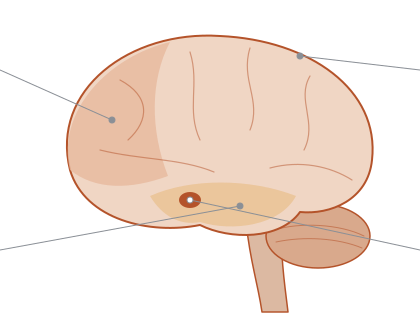
Larger brains in some young children Established
On average, young autistic children, mostly boys in these studies, have larger brains than non-autistic children of the same age. The difference shows up on brain scans by about age 2 to 2½.1,2 In one large group of children followed in California, the average difference came from a subgroup: autistic boys with typical-sized brains did not differ from other boys, and autistic girls' brains were not larger at all.3
Pooling dozens of studies, about 1 in 6 autistic people (15.7%) have an unusually large head, and about 1 in 11 (9.1%) an unusually large brain.4 The usual definition of a large head (at or above the 97th percentile, or 2 standard deviations above average) means about 2 to 3% of any reference group counts as having one.5 The brain-size difference in this meta-analysis was larger in autistic people it described as "low functioning".4
Scientists use brain scans to measure how big a brain is. On average, young autistic kids have bigger brains than other kids their age.1,2 This shows up by about age 2. Most of these kids were boys.
In one big study, the difference came from a small group of autistic boys. The other autistic boys had brains of usual size. Autistic girls did not have bigger brains.3
About 1 in 6 autistic people have a large head. About 1 in 11 have a large brain.4
Several MRI studies that followed the same children agree that brains are larger on average at ages 2 to 4. The samples were mostly boys. Head-size studies are less consistent (see the note below).
Many early studies compared autistic children with national growth charts. Studies that measured local children for comparison, and one study of every child in a region of Israel, found little or no difference in head size.6,7 In one large study of families, 14.7% of autistic children had a large head by standard charts, and so did 12.7 to 19.6% of their non-autistic relatives. Measured against what their parents' heads predicted, 3.6% of the autistic children did.5
A scanner that uses magnets to take detailed pictures of the brain without surgery.
Many studies agree on this. Most of them looked at boys.
A scanner that uses strong magnets to take pictures of the inside of the brain.
What grows, and when Emerging
The extra size seems to lie mostly in the area of the cortex rather than in its thickness.1,8 A third study, of 3-year-old autistic boys, found the same pattern, mainly in the boys whose brains were unusually large for their height.9
In babies with an autistic older brother or sister, the 15 who were later diagnosed showed the surface of the cortex spreading faster between 6 and 12 months. Faster growth in total brain size followed between 12 and 24 months.8 That faster second-year growth went, weakly, with more marked social differences at age 2.8
Which regions grow most is less clear: one study found the biggest differences in frontal, temporal and cingulate cortex; another found temporal white matter enlarged.2,1 Some studies report faster growth of the amygdala: between 6 and 24 months in infants later diagnosed, and between about 3 and 4 years in autistic boys, with wide variation between children.10,11 Across all ages, the largest study found the amygdala slightly smaller on average in autistic people, so the early findings and the later averages point in different directions.12
In two separate groups of infants with an autistic older sibling, those later diagnosed had more fluid around the brain from about 6 months, and more fluid went with more marked autistic traits.13,14 A third study from the same research group found the same excess at ages 2 to 4, including in children with no autistic sibling.15
The extra size seems to come from the brain's outer layer spreading out. It does not seem to get thicker.1,8,9
One study followed babies who had an autistic big brother or sister. Fifteen of these babies were later found to be autistic. Their brain's outer layer spread out faster before their first birthday.8 In their second year, the whole brain grew faster. That faster growth went, a little, with bigger social differences at age 2.8
Scientists do not agree yet on which parts grow most.2,1 Some studies say the amygdala grows faster in early childhood. Kids were very different from each other.10,11 Later in life, the amygdala is a little smaller on average.12
Some babies who were later found to be autistic had more fluid around the brain. This showed up by about 6 months.13,14,15
Most of these findings come from two research groups and from infants with an autistic older sibling. Few have been repeated by a fully independent team.
The folded outer layer of the brain, a few millimetres thick.
A small almond-shaped structure deep in the brain, involved in emotion and in noticing what matters.
The clear fluid between the brain and the skull.
Each finding comes from one team or one small group of kids. Nobody else has checked it yet.
The wrinkly outer layer of the brain.
A small part deep in the brain. It helps with feelings and noticing what matters.
The clear liquid between the brain and the skull.
A subgroup with large brains Emerging
In one long-running study, about 15% of autistic boys had brains large for their body size at age 3. Their heads were already larger by about age 2, though they had not been at birth.16 In this subgroup the enlargement lasted through about age 11, with no sign of shrinking back.3
Some studies report bigger brain-size differences, or faster early head growth, in autistic children who also have an intellectual disability.17,4 The same California study found no difference in early developmental scores between boys with and without enlarged brains.16
Changes in single genes account for a few cases. In one small study, 3 of 18 autistic children with very large heads carried a change in the PTEN gene.18 In a study of 3,730 children, those with changes in the CHD8 gene often had a large head.19
In one long study, about 15 out of every 100 autistic boys had big brains for their body size. Their heads were normal at birth. By age 2, their heads were bigger.16 Their brains stayed bigger until at least age 11.3
Some studies found bigger differences in kids who also have an intellectual disability.17,4 But the California study found no difference in early test scores.16
A few kids have a change in one gene that makes their head and brain bigger.18,19
These findings come mainly from one long-running study of boys in California.
Most of this comes from one long study of boys in California.
When learning and everyday thinking skills develop much more slowly than usual.
Girls and boys Emerging
Most brain-growth studies have included mainly males.3,20 Two long-running studies covering ages 3 to 36 were all male, and one adult study was about 90% men.21,22,23
Where girls were studied, autistic girls' brains were not larger on average but grew more slowly between about ages 3 and 5½.3 A 2024 study found autistic girls had thicker cortex around age 3 and faster thinning afterwards, across a wider area than in autistic boys. The differences had faded by about age 11½.20 Both of these findings come from the same group of children in California.
Studies of adults and of large multi-site samples also find that some brain differences linked to autism depend on sex.24,25
Most brain growth studies have looked mainly at boys.3,20 Some long studies included no girls at all.21,22,23
Autistic girls did not have bigger brains. Their brains grew a bit more slowly between ages 3 and 5.3 One study found that autistic girls had a thicker outer layer at age 3. It then got thinner faster. By about age 11, the differences had mostly faded.20 Both of these findings come from the same group of kids.3
Single studies, not yet repeated. Most brain-growth research has included mainly boys.
Each finding comes from one study. Most brain studies have been about boys.
Childhood and adolescence Emerging
Whether early enlargement "evens out" later is unsettled. One study of males aged 6 to 35 found total brain size meeting the typical curve between 10 and 15, though some regions kept changing atypically into adulthood.21 Another study found that boys in the large-brain subgroup kept their larger brains through about age 11.3 Its authors argue that studies comparing different age groups may miss people with large brains in their older samples, making it look as if the difference disappears.3
In the largest study, of about 1,600 autistic people aged 2 to 64 scanned once each, the cortex was slightly thicker in frontal regions and slightly thinner in temporal regions. The differences were very small, so autistic and non-autistic people overlapped heavily, and they were largest around adolescence.12 An analysis of about 1,000 people aged 6 to 35 found only a few small differences in brain anatomy.26
Studies of how the cortex thins disagree. Two that tracked people over time report faster thinning in autistic adolescents: one in just two left-side regions, the other followed by slower-than-typical thinning in early adulthood.27,22 One study comparing ages 7 to 39 found less thinning with age, and another, of adults, found more.28,23
Does the extra size even out as kids grow up? Scientists are not sure yet. One study found brain size became usual between ages 10 and 15.21 Another study found that boys with big brains kept them until at least age 11.3 Its authors think some studies miss older people with big brains. That could make the difference look like it goes away.3
The biggest study scanned about 1,600 autistic people once each. The front of the brain's outer layer was a little thicker. The sides were a little thinner. The differences were very small. Most autistic and non-autistic people overlapped.12,26
Studies disagree about how the outer layer thins with age. Some found faster thinning in autistic teenagers.27,22 Others found less thinning, or more thinning in adults.28,23
Studies disagree on direction, and the ones that followed people over time were small or all male.
Studies disagree, and the ones that followed kids over time were small.
Every brain differs Established
When each autistic person's brain is compared with what is typical for their age, studies of children and young adults, mostly male, find large and varied individual differences: two autistic people rarely differ from the typical range in the same places.29,30 Group-average differences in these studies range from few and small to present in some brain measures.29,31
A 2026 study made a related point for brain connectivity. Using normative models on resting-state scans from 1,824 people (796 autistic, ages 5 to 58, across 32 sites), it found that no single connection was extremely atypical in more than about 4% of people in either group, so a group difference on any single connection was not typical of the group. The deviations were not random, though. They converged: in the autistic group they overlapped up to twice as much as in the non-autistic group, and they clustered in the same regions and networks. Connectivity was weaker in sensorimotor and attention areas and stronger involving the frontoparietal and default mode networks and their links with the rest of the brain. The deviation patterns went with social and cognitive scores.43 The autistic group was about 82% male, and the method detected deviations less well at the youngest and oldest ages.
Scientists can compare each autistic person's brain with what is usual for their age. When they do, every brain is different in its own way. Two autistic people rarely differ in the same places.29,30 Studies disagree about how big the average differences are.29,31
A newer study checked this in 1,824 people. It found that no single link between brain areas was very unusual in more than about 4 in 100 people. But the unusual links were not random. They showed up in the same places more often in autistic people than in others. Some areas had weaker links, and some had stronger links.43
Two independent studies using the same method agree on large individual differences. One of them, and a related study, included only males. A third, larger study using the same method agrees, and finds the differences overlap more in autistic people than in non-autistic people; it re-analyzes shared public datasets (ABIDE and EU-AIMS LEAP) rather than a new sample. How big the group-average differences are varies between studies.
A study of six conditions, including autism, found that fewer than 7% of people with the same diagnosis had an extreme difference in the same brain region (under 5% for autism, in an all-male autism sample). The differences often fell within shared brain networks.32
Separate studies using the same method agree on this. Most of the people studied were boys and men. A newer, bigger study agrees.
A study of six conditions, including autism, found that fewer than 7% of people with the same diagnosis had an extreme difference in the same brain region (under 5% for autism, in an all-male autism sample). The differences often fell within shared brain networks.32
Adulthood and aging Emerging
One study of 157 autistic adults (ages 18 to 64, about 90% men, median age 26) found a stronger link between older age and thinner cortex than in non-autistic adults, mostly in the left temporal lobe.23 Each person was scanned once, so the result shows that older participants had thinner cortex. It cannot show anyone's cortex thinning over time.
A study of 51 autistic adults aged 30 to 75, all with an IQ above 80, found no difference in cortical volume, thickness or surface area.33 A small study of 29 autistic adults aged 40 to 70 found that tracts linking deep brain structures to the cortex changed with age in parallel with non-autistic adults, not faster, though some were smaller in the autistic group.34
As of October 2026, repeated brain scans of autistic people stop at around age 35 to 39. A study of adults aged 40 to 70 repeated memory tests over about two years but scanned the brain only once.21,22,35 The adult studies included few or no autistic people with an intellectual disability, so their findings may not apply to them.23,33
One study scanned 157 autistic adults once each. Most were young men. The older adults had a thinner outer layer than older non-autistic adults.23 This does not show anyone's brain changing over time.
A study of 51 autistic adults aged 30 to 75 found no differences.33 A small study of adults aged 40 to 70 found some brain pathways changing with age at the usual speed.34
In the studies we looked at, repeated brain scans of autistic people stop at about age 39.21,22,35 Most of the adults studied did not have an intellectual disability. So the results may not apply to everyone.23,33
All of the adult studies scanned people once. None has followed the same autistic adults' brains into later life.
A study that measures different people at different ages once, rather than following the same people as they age.
Each adult was scanned only once. No study has followed the same adults' brains as they got older.
A study that measures different people once, instead of following the same people over time.
What might drive early growth Hypothesis
It has been proposed that early overgrowth starts before birth, from the brain making too many cells.36,37 One small preliminary study of the brains of 7 autistic boys who had died (compared with 6 non-autistic boys) reported about two-thirds more neurons in the prefrontal cortex.38
Brain organoids grown from the cells of 10 autistic toddlers were about 40% larger than those from 6 non-autistic toddlers, in two separate batches, and larger size went with more marked social differences. This is an association in 16 donors and has not been repeated by another team.37 Stem cells from 8 autistic people with enlarged brains multiplied faster in the lab,39 and organoids from 4 autistic people with large heads made too many inhibitory neurons.40
Not every finding fits. A study of the brains of 9 autistic males aged about 10 to their mid-40s found fewer neurons in the amygdala, not more.41 At most, this explanation applies to the subgroup with large brains, not to autistic people in general.
A separate proposal concerns pruning, the loss of surplus connections between nerve cells during childhood and adolescence. In one post-mortem study of 10 autistic and 10 non-autistic brains aged about 2 to 20, the number of dendritic spines fell by about 16% between childhood and adolescence in the autistic tissue, against about 41 to 45% in the non-autistic tissue.42 Whether this connects to the cortical thickness changes described above is an open question.
Some scientists think the extra growth starts before birth. The idea is that the brain makes too many cells.36,37 One small study looked at the brains of 7 autistic boys who had died. It found about two-thirds more nerve cells in the front of the brain.38
Scientists also grew tiny brain-like clumps from the cells of 10 autistic toddlers. These clumps grew bigger than ones from other toddlers. Only 16 people were tested.37 Cells from other autistic people with big heads also grew faster.39,40
Not every clue fits. One study found fewer nerve cells in one part of the brain, not more.41
Another idea is about pruning. As kids grow, the brain clears away extra links between nerve cells. One small study found fewer links cleared away in autistic brains.42
A proposed explanation for the subgroup with large brains. It rests on small studies of brains after death and of cells grown in a lab, and has not been tested in living children.
A tiny cluster of brain-like tissue grown in a dish from a person's own cells.
This is an idea that fits some clues. It might explain the kids with big brains, not autism in general.
A tiny clump of brain-like cells grown in a dish.
Sources
- 1Hazlett HC, Poe MD, Gerig G, et al. Early brain overgrowth in autism associated with an increase in cortical surface area before age 2 years. Archives of General Psychiatry 2011. https://doi.org/10.1001/archgenpsychiatry.2011.39 ; https://pubmed.ncbi.nlm.nih.gov/21536976/
- 2Schumann CM, Bloss CS, Barnes CC, et al. Longitudinal magnetic resonance imaging study of cortical development through early childhood in autism. Journal of Neuroscience 2010. https://doi.org/10.1523/JNEUROSCI.5714-09.2010 ; https://pubmed.ncbi.nlm.nih.gov/20335478/
- 3Lee JK, Andrews DS, Ozonoff S, et al. Longitudinal evaluation of cerebral growth across childhood in boys and girls with autism spectrum disorder. Biological Psychiatry 2021;90:286–294. https://doi.org/10.1016/j.biopsych.2020.10.014 ; https://pubmed.ncbi.nlm.nih.gov/33388135/
- 4Sacco R, Gabriele S, Persico AM. Head circumference and brain size in autism spectrum disorder: a systematic review and meta-analysis. Psychiatry Research: Neuroimaging 2015. https://doi.org/10.1016/j.pscychresns.2015.08.016 ; https://pubmed.ncbi.nlm.nih.gov/26456415/
- 5Chaste P, Klei L, Sanders SJ, et al. Adjusting Head Circumference for Covariates in Autism: Clinical Correlates of a Highly Heritable Continuous Trait. Biological Psychiatry 2013. https://doi.org/10.1016/j.biopsych.2013.04.018
- 6Raznahan A, Wallace GL, Antezana L, et al. Compared to what? Early brain overgrowth in autism and the perils of population norms. Biological Psychiatry 2013;74:563–575. https://doi.org/10.1016/j.biopsych.2013.03.022 ; https://pubmed.ncbi.nlm.nih.gov/23706681/
- 7Dinstein I, Haar S, Atsmon S, Schtaerman H. No evidence of early head circumference enlargements in children later diagnosed with autism in Israel. Molecular Autism 2017. https://doi.org/10.1186/s13229-017-0129-9 ; https://pubmed.ncbi.nlm.nih.gov/28344758/
- 8Hazlett HC, Gu H, Munsell BC, et al. Early brain development in infants at high risk for autism spectrum disorder. Nature 2017;542:348–351. https://doi.org/10.1038/nature21369 ; https://pubmed.ncbi.nlm.nih.gov/28202961/
- 9Ohta H, Nordahl CW, Iosif A, et al. Increased Surface Area, but not Cortical Thickness, in a Subset of Young Boys With Autism Spectrum Disorder. Autism Research 2016. https://doi.org/10.1002/aur.1520
- 10Shen MD, Swanson MR, Wolff JJ, et al. Subcortical brain development in autism and fragile X syndrome: evidence for dynamic, age- and disorder-specific trajectories in infancy. American Journal of Psychiatry 2022. https://doi.org/10.1176/appi.ajp.21090896 ; https://pubmed.ncbi.nlm.nih.gov/35331012/
- 11Nordahl CW, Scholz R, Yang X, et al. Increased rate of amygdala growth in children aged 2 to 4 years with autism spectrum disorders: a longitudinal study. Archives of General Psychiatry 2012. https://doi.org/10.1001/archgenpsychiatry.2011.145 ; https://pubmed.ncbi.nlm.nih.gov/22213789/
- 12van Rooij D, Anagnostou E, Arango C, et al. Cortical and subcortical brain morphometry differences between patients with autism spectrum disorder and healthy individuals across the lifespan: results from the ENIGMA ASD Working Group. American Journal of Psychiatry 2018;175:359–369. https://doi.org/10.1176/appi.ajp.2017.17010100 ; https://pubmed.ncbi.nlm.nih.gov/29145754/
- 13Shen MD, Nordahl CW, Young GS, et al. Early brain enlargement and elevated extra-axial fluid in infants who develop autism spectrum disorder. Brain 2013. https://doi.org/10.1093/brain/awt166
- 14Shen MD, Kim SH, McKinstry RC, et al. Increased Extra-axial Cerebrospinal Fluid in High-Risk Infants Who Later Develop Autism. Biological Psychiatry 2017. https://doi.org/10.1016/j.biopsych.2017.02.1095
- 15Shen MD, Nordahl CW, Li DD, et al. Extra-axial cerebrospinal fluid in high-risk and normal-risk children with autism aged 2–4 years: a case-control study. The Lancet Psychiatry 2018. https://doi.org/10.1016/s2215-0366(18)30294-3
- 16Libero LE, Nordahl CW, Li DD, et al. Persistence of megalencephaly in a subgroup of young boys with autism spectrum disorder. Autism Research 2016. https://doi.org/10.1002/aur.1643 ; https://pubmed.ncbi.nlm.nih.gov/27273931/
- 17McKeague IW, Brown AS, Bao Y, et al. Autism with intellectual disability related to dynamics of head circumference growth during early infancy. Biological Psychiatry 2015. https://doi.org/10.1016/j.biopsych.2014.08.008 ; https://pubmed.ncbi.nlm.nih.gov/25444163/
- 18Butler MG, Dasouki MJ, Zhou XP, et al. Subset of individuals with autism spectrum disorders and extreme macrocephaly associated with germline PTEN tumour suppressor gene mutations. Journal of Medical Genetics 2005. https://doi.org/10.1136/jmg.2004.024646
- 19Bernier R, Golzio C, Xiong B, et al. Disruptive CHD8 Mutations Define a Subtype of Autism Early in Development. Cell 2014. https://doi.org/10.1016/j.cell.2014.06.017
- 20Andrews DS, Diers K, Lee JK, et al. Sex differences in trajectories of cortical development in autistic children from 2–13 years of age. Molecular Psychiatry 2024. https://doi.org/10.1038/s41380-024-02592-8 ; https://pubmed.ncbi.nlm.nih.gov/38755243/
- 21Lange N, Travers BG, Bigler ED, et al. Longitudinal volumetric brain changes in autism spectrum disorder ages 6–35 years. Autism Research 2015. https://doi.org/10.1002/aur.1427 ; https://pubmed.ncbi.nlm.nih.gov/25381736/
- 22Zielinski BA, Prigge MB, Nielsen JA, et al. Longitudinal changes in cortical thickness in autism and typical development. Brain 2014;137:1799–1812. https://doi.org/10.1093/brain/awu083 ; https://pmc.ncbi.nlm.nih.gov/articles/PMC4032101/
- 23Braden BB, Riecken C. Thinning faster? Age-related cortical thickness differences in adults with autism spectrum disorder. Research in Autism Spectrum Disorders 2019. https://pmc.ncbi.nlm.nih.gov/articles/PMC7304569/
- 24Lai MC, Lombardo MV, Suckling J, et al. Biological sex affects the neurobiology of autism. Brain 2013. https://doi.org/10.1093/brain/awt216
- 25Bedford SA, Park MTM, Devenyi GA, et al. Large-scale analyses of the relationship between sex, age and intelligence quotient heterogeneity and cortical morphometry in autism spectrum disorder. Molecular Psychiatry 2020. https://doi.org/10.1038/s41380-019-0420-6
- 26Haar S, Berman S, Behrmann M, et al. Anatomical Abnormalities in Autism?. Cerebral Cortex 2016. https://doi.org/10.1093/cercor/bhu242
- 27Wallace GL, Eisenberg IW, Robustelli B, et al. Longitudinal cortical development during adolescence and young adulthood in autism spectrum disorder: increased cortical thinning but comparable surface area changes. Journal of the American Academy of Child and Adolescent Psychiatry 2015;54:464–469. https://doi.org/10.1016/j.jaac.2015.03.007 ; https://pubmed.ncbi.nlm.nih.gov/26004661/
- 28Doyle-Thomas KAR, Duerden EG, Taylor MJ, et al. Effects of age and symptomatology on cortical thickness in autism spectrum disorders. Research in Autism Spectrum Disorders 2013. https://pmc.ncbi.nlm.nih.gov/articles/PMC3652338/
- 29Zabihi M, Oldehinkel M, Wolfers T, et al. Dissecting the heterogeneous cortical anatomy of autism spectrum disorder using normative models. Biological Psychiatry: Cognitive Neuroscience and Neuroimaging 2019. https://doi.org/10.1016/j.bpsc.2018.11.013 ; https://pubmed.ncbi.nlm.nih.gov/30799285/
- 30Bethlehem RAI, Seidlitz J, Romero-Garcia R, et al. A normative modelling approach reveals age-atypical cortical thickness in a subgroup of males with autism spectrum disorder. Communications Biology 2020. https://doi.org/10.1038/s42003-020-01212-9 ; https://pubmed.ncbi.nlm.nih.gov/32887930/
- 31Shan X, Uddin LQ, Xiao J, et al. Mapping the heterogeneous brain structural phenotype of autism spectrum disorder using the normative model. Biological Psychiatry 2022. https://doi.org/10.1016/j.biopsych.2022.01.011 ; https://pubmed.ncbi.nlm.nih.gov/35367047/
- 32Segal A, Parkes L, Aquino K, et al. Regional, circuit and network heterogeneity of brain abnormalities in psychiatric disorders. Nature Neuroscience 2023. https://doi.org/10.1038/s41593-023-01404-6 ; https://pubmed.ncbi.nlm.nih.gov/37580620/
- 33Koolschijn PCMP, Geurts HM. Gray matter characteristics in mid and old aged adults with ASD. Journal of Autism and Developmental Disorders 2016. https://doi.org/10.1007/s10803-016-2810-9 ; https://pubmed.ncbi.nlm.nih.gov/27177894/
- 34Cordova M, Hau J, Schadler A, et al. Structure of subcortico-cortical tracts in middle-aged and older adults with autism spectrum disorder. Cerebral Cortex 2024. https://doi.org/10.1093/cercor/bhae457 ; https://pubmed.ncbi.nlm.nih.gov/39707985/
- 35Walsh MJM, Ofori E, Pagni BA, et al. Preliminary findings of accelerated visual memory decline and baseline brain correlates in middle-age and older adults with autism: the case for hippocampal free-water. Frontiers in Aging Neuroscience 2022. https://doi.org/10.3389/fnagi.2022.1029166 ; https://pubmed.ncbi.nlm.nih.gov/36437999/
- 36Courchesne E, Campbell K, Solso S. Brain growth across the life span in autism: age-specific changes in anatomical pathology. Brain Research 2011;1380:138–145. https://doi.org/10.1016/j.brainres.2010.09.101 ; https://pmc.ncbi.nlm.nih.gov/articles/PMC4500507/
- 37Courchesne E, Taluja V, Nazari S, et al. Embryonic origin of two ASD subtypes of social symptom severity: the larger the brain cortical organoid size, the more severe the social symptoms. Molecular Autism 2024. https://doi.org/10.1186/s13229-024-00602-8 ; https://pubmed.ncbi.nlm.nih.gov/38790065/
- 38Courchesne E, Mouton PR, Calhoun ME, et al. Neuron number and size in prefrontal cortex of children with autism. JAMA 2011;306:2001–2010. https://doi.org/10.1001/jama.2011.1638 ; https://pubmed.ncbi.nlm.nih.gov/22068992/
- 39Marchetto MC, Belinson H, Tian Y, et al. Altered proliferation and networks in neural cells derived from idiopathic autistic individuals. Molecular Psychiatry 2017. https://doi.org/10.1038/mp.2016.95
- 40Mariani J, Coppola G, Zhang P, et al. FOXG1-Dependent Dysregulation of GABA/Glutamate Neuron Differentiation in Autism Spectrum Disorders. Cell 2015. https://doi.org/10.1016/j.cell.2015.06.034
- 41Schumann CM, Amaral DG. Stereological Analysis of Amygdala Neuron Number in Autism. The Journal of Neuroscience 2006. https://doi.org/10.1523/jneurosci.1285-06.2006
- 42Tang G, Gudsnuk K, Kuo SH, et al. Loss of mTOR-dependent macroautophagy causes autistic-like synaptic pruning deficits. Neuron, 2014. https://doi.org/10.1016/j.neuron.2014.07.040
- 43Ilioska I, Oldehinkel M, Llera A, et al. Multiscale heterogeneity of atypical functional connectivity in autism. Nature Mental Health, 2026;4(6):1010–1020. https://doi.org/10.1038/s44220-026-00656-y